 By
By
- Samuel Boyd, M.D., Editor in Chief and Executive Vice President, HIGHLIGHTS OF OPHTHALMOLOGY., Di rector, La ser Section and Associa te Direc tor, Retina and Vitreous Department, , Clinica Bo yd – Ophthal mo lo gy Center, Panama, Rep. of Panama
- Rosario Brancato, M.D., Professor of Ophthalmology , Director of the School of Ophthalmologic Specialization, San Raffaele University Hospital, Milan, Italy
- Bradley Straatsma, M.D., Professor and Director Emeritus, Jules Stein Eye Institute, University of California, Los Angeles (UCLA), Los Angeles, California, U.S.A.
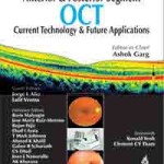
Optical Coherence Tomography (OCT) is a technology that has come of age. From a laboratory tool for performing a single A-scan, to a research device for creating cross-sectional images of ocular tissues in vivo, OCT has evolved into a clinical instrument that is used to detect and direct the treatment of glaucoma, retinal diseases and anterior segment pathologies. This technology continues to evolve, and the current volume explores current and future uses for OCT. The current compilation covers the use and interpretation of OCT, the applications of OCT in ophthalmology, including uses in the anterior segment and retina. OCT in the diagnosis and management of glaucoma and age related macular degeneration is described. In addition, the integration of OCT and fluorescein angiography is discussed, as well as the use of OCT in the diagnosis and management of ocular tumors. Newer iterations of OCT permit three dimensional imaging, due to advances that allow very high speed, high resolution capture of images. Instead of 400 A-scans per second that were possible with conventional, or time-domain OCT, spectral domain OCT enables the acquisition of 20,000 – 40,000 A-scans per second. These three dimensional OCT images present opportunities and challenges. Improvements in resolution may lead to increased reproducibility, sensitivity and specificity. Three dimensional OCT (3D OCT) permits the creation of an OCT fundus image, which facilitates precise image registration. The higher resolution allows segmentation and mapping of intraretinal layers. Newer versions of OCT improve visualization of tissues relevant in the detection and management of ocular disease. On the other hand, 3D OCT creates very large data sets, and optimal methods for analysis are still in the process of being created. OCT is a robust technology that continues to evolve. OCT is a useful tool for diagnosis and management of eye diseases and may provide a cost effective diagnostic utility for basic and clinical studies of eye diseases. Of course, the ultimate goal is the earliest possible detection of a disease and its progression in order to prevent visual loss in our patients.


